MRI-conditional Extracorporeal Membrane Oxygenator
Congenital heart disease (CHD) describes one of the most common birth defects in humans with a prevalence of around 7.2 per 1’000 births in Europe. Around 7% of newborn with CHD require immediate open-heart surgery within the first 6 months of their life. During these surgical interventions, a cardiopulmonary bypass (CPB) is used to sustain circulation and oxygenation of the blood. However, the operating point of these CPBs has not yet been determined sufficiently in regards of minimizing irreversible brain damage during surgery. A promising approach to gain more insights into these dynamics would be to use magnetic resonance imaging (MRI) while the CPB is attached.
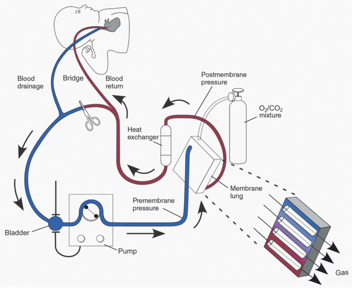
A lot of restrictions exist for devices to be used in an MRI environment, such as material constraints. Possible solutions for these issues were to use long blood tubes or long drive shafts. Using long blood tubes is not feasible with young children, since the drained blood volume would exceed the blood volume of their bodies. Long drive shafts on the other hand are very fragile and impractical.
This project is conducted in cooperation between ETH Zurich and the University Hospital Zurich and aims to develop a fully MRI conditional extra-corporeal membrane oxygenation device. This device should fulfill the most important roles of conventional heart-lung machines such as maintaining blood circulation and oxygenation.