Cardex
Many catheter-based procedures on the heart require access to the left atrium. At birth a passage through the interatrial septum exists. However, this passage closes off in later stages of growth. To re-establish this connection, a needle of approx. 90cm length is inserted at the groin. The device is advanced through the inferior vena cava up to the right atrium, to puncture the interatrial septum. The surgeon has no direct vision and therefore guides the needle with the use of fluoroscopy and echocardiography as imaging technologies. This whole procedure is called "Transseptal Puncture Procedure".
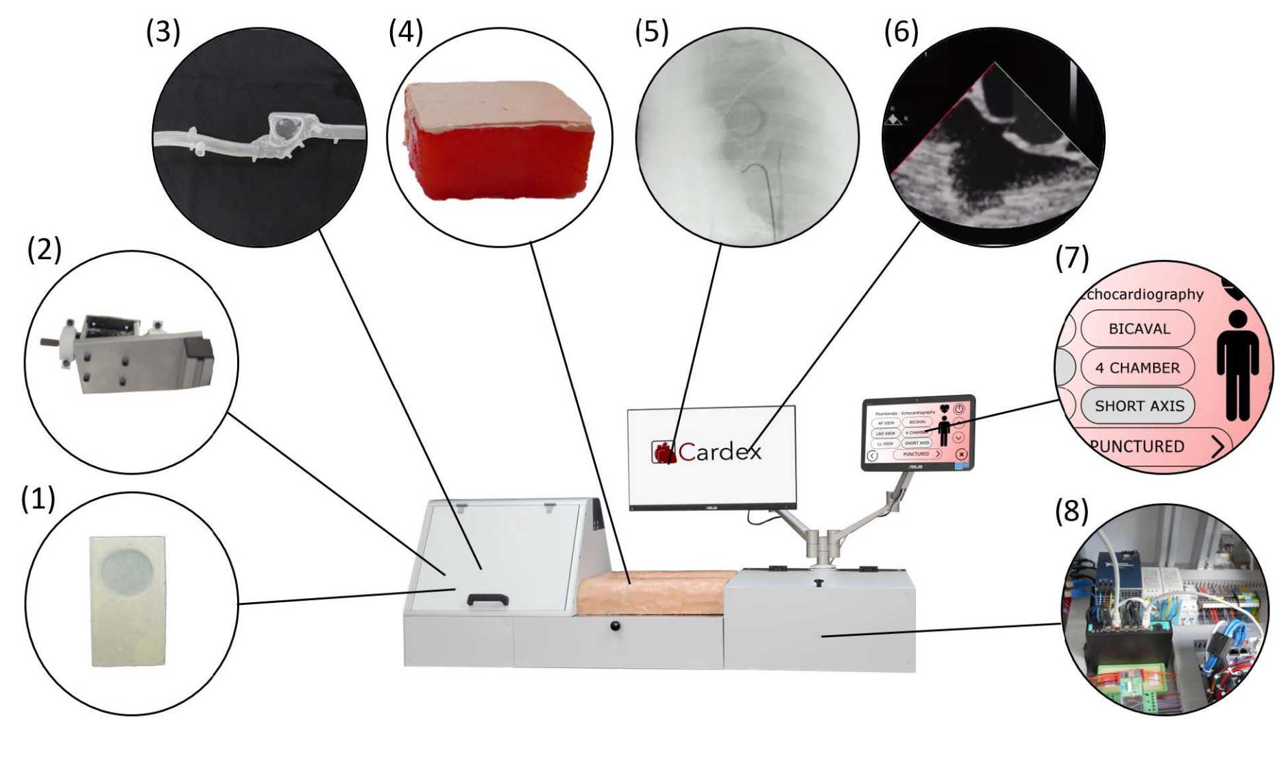
Our goal was to construct a simulator for this procedure. By providing realistic handling in combination with direct feedback, this novel experience offers a better preparation for transseptal puncture. There are already simulators trying to tackle this subject but most of these products are insufficient solutions. Either they are too far from reality or they require animal hearts and extensive preparations. That is why we developed a high-fidelity simulator seen in Fig. 1, which can be used for training and qualification of medical students and practicing doctors, in order to optimally prepare them for the actual surgery.
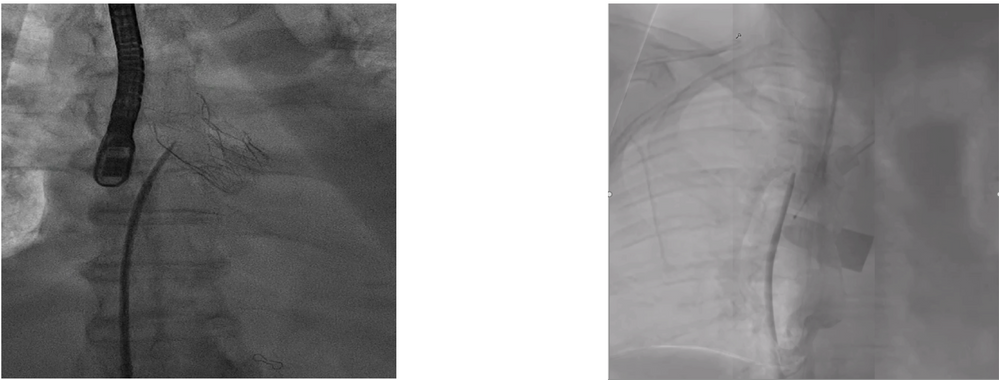
One of the main features of the simulator is the mimicing of X-Ray imaging. Real X-Ray as used in the operation theatre emits radiation, which is harmful for both, the surgeon and the patient. In Fig. 2 a image of the simulated X-Ray can be seen next to a real X-Ray image. This is without ever exposing the learning surgeon to harmful radiation.
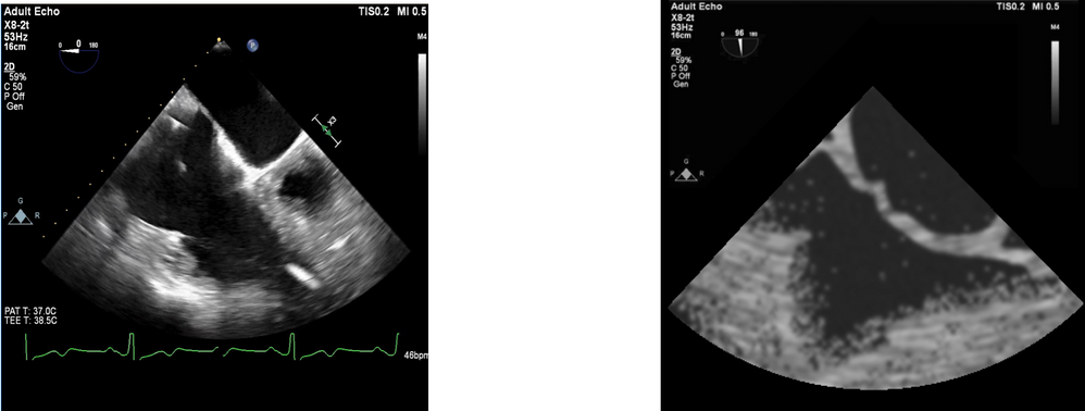
Similarly the mimicing of echocardiography is crucial for the transseptal puncture procedure. The simulation of the ultrasound image was achieved by a scanning system based on a stereo camera setup, that constantly measures the deformation of the Fossa Ovalis and transforms this information into a classical ultrasound image. Fig. 3 shows the comparison between a real ultrasound image and a simulated one.
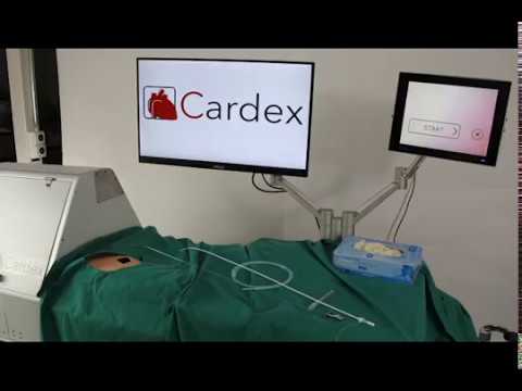
There are many other engineering solutions, as can be seen in the system overview in Fig. 1, that contribute to the performance of this novel simulator. The following video shows all the steps of the whole transseptal puncture procedure and performed on the simulator.
Impressions and related material: